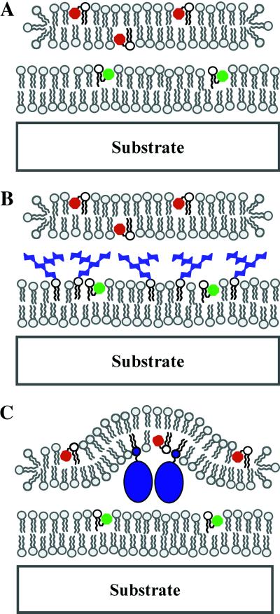
Fig 1.
Schematic diagrams of supported intermembrane junctions. Molecular species on the membrane surface influence intermembrane spacing. A and B depict situations corresponding to the PI- and GM1-containing membrane junctions described in Results and Discussion. C illustrates how membrane-bending deformations can couple to molecular organization in the junction, which is representative of the PEG-lipid junctions discussed later in Results and Discussion.

