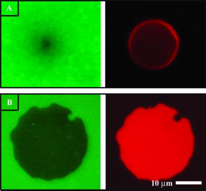
Fig 2.
(A) Fluorescence image of the lower (green, Left) and upper (red, Right) membranes of an unruptured GUV on a supported membrane. (B) Image of a supported membrane junction formed after rupture of a GUV. In both cases, patterns seen in the lower membranes result from intermembrane FRET to acceptors in the upper membrane.

