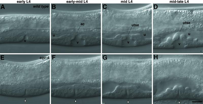
Fig 1.
Nomarski photomicrographs of vulval morphogenesis. Vulval morphogenesis in WT animals (A–D) and sqv-4(n2827) mutants (E–H) in sequential developmental stages. Anterior is on the left, and dorsal is up. (A) The vulval extracellular space (v) is a clear area devoid of granules. (B) The vulval extracellular space (v) is bottle-shaped. The uterine extracellular space (u) is a narrow area devoid of granules spanning the anterior to posterior ends and is separated from the vulval extracellular space by the anchor cell (ac). (C) The vulval (v) and uterine (u) extracellular spaces are separated by a thin planar cytoplasmic process of a uterine cell (utse). (D) A vulval extracellular space with an expanded dorsal end causing the utse cytoplasmic process to bend inward. E–H approximately correspond in developmental stage to the WT animals in A–D, respectively. The white arrowhead indicates the vulval extracellular space. (Scale bar = 10 μm.)

