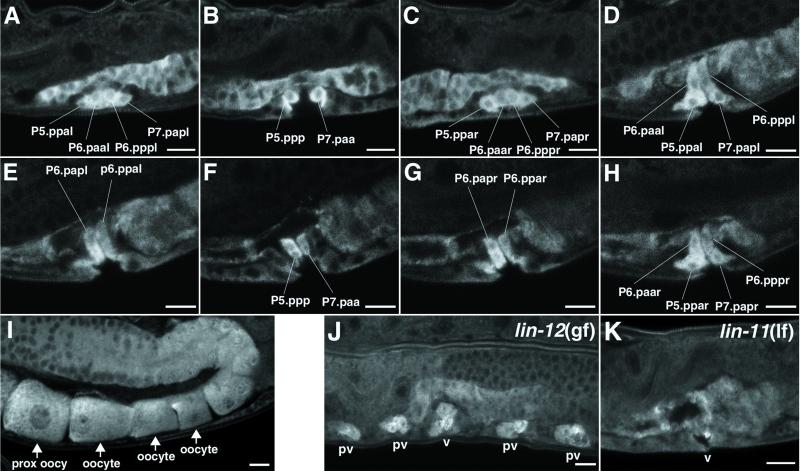
Fig 3.
SQV-4 protein expression. (A–H) Selected confocal images of SQV-4 Ab staining in multiple planes of focus along the left-to-right axis of the WT animal. Vulval cells with increased SQV-4 expression are indicated. (A–C) SQV-4 expression is increased in cells containing 10 of 22 vulval nuclei in the early L4 stage. The cells strongly stained with SQV-4 Abs dorsal to the vulval cells are in the uterus. The images are arranged from the left-most plane of focus to the right-most plane. (A) Vulval cells containing four nuclei (P5.ppal, P6.paal, P6.pppl, and P7.papl) on the left side of the worm. (B) Two vulval cells (P5.ppp and P7.paa) in the middle plane of the worm. (C) Vulval cells containing four nuclei (P5.ppar, P6.paar, P6.pppr, and P7.papr) on the right side of the worm. (D–H) SQV-4 expression is increased in cells containing 14 of 22 vulval nuclei in the mid-late L4 stage. The images are arranged from the left-most plane of focus to the right-most plane. (D) Vulval cells containing the same four nuclei as in A. (E) Vulval cells containing two of the four dorsal-most nuclei (P6.papl and P6.ppal), which were not stained at an earlier stage. (F) Vulval cells containing the same two nuclei as in B. (G) Vulval cells containing two of the four dorsal-most nuclei (P6.papr and P6.ppar), which were not stained at an earlier stage. (H) Vulval cells containing the same four nuclei as in C. (I) SQV-4 Abs stain oocytes. A row of oocytes in an adult hermaphrodite stained with SQV-4 Abs is indicated with arrows. The oocyte most proximal to the uterus is located at the lower left. (J–K) SQV-4 protein expression in mutants with abnormal vulval development. (J) Confocal image of a lin-12(gf) mutant fixed and stained with SQV-4 Abs. Each pseudovulva (pv) has three nuclei strongly stained with SQV-4 Abs. The functional vulva (v) has six nuclei strongly stained with SQV-4 Abs. Not all staining is shown here. (K) Confocal image of an L4 lin-11(lf) mutant fixed and stained with SQV-4 Abs. Staining by SQV-4 Abs is limited to one cell in this focal plane. Staining is absent in vulval cells in other focal planes (not shown). (Scale bar = 10 μm.)

