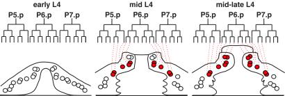
Fig 4.
Diagram of SQV-4 expression in vulval cells. WT vulval cell lineages and lateral views of vulva at three developmental stages. The vulva on the left, center, and right approximate the vulva in Fig. 1 A, C, and D, respectively. Circles indicate vulval cell nuclei, and overlapping circles indicate nuclei that are in different planes of focus along the left–right axis. Red circles, nuclei of the cells with increased SQV-4 expression. Open circles, nuclei of cells without detectable SQV-4 expression.

