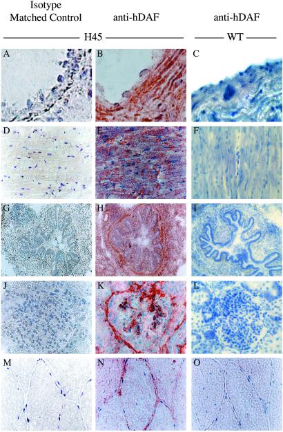
Fig 5.
Immunostaining for hDAF on frozen tissue sections from transgenic pigs. Serial sections of aorta (A and B), heart (D and E), lung (G and H), kidney (J and K), and muscle (M and N) from transgenic pig H45 were stained with IF7 anti-hDAF mAb (B, E, H, K, and N) or an isotype matched control (A, D, G, J, and M). The aortic wall shows strong positivity mostly in the extracellular matrix. The heart shows hDAF expression mostly in the plasmalemma of the muscle fibers. In the lung, the protein is expressed at a very high level in all tissues. The vascular tuft of the glomeruli and the vascular spaces of the Bowman capsule of the kidney show strong positivity. In skeletal muscle, hDAF was expressed in the sarcoplasm and on the sarcolemma of fibers in a peripheral location. Sections of aorta, heart, lung, kidney, and muscle from a control pig were stained with the same anti-hDAF mAb (C, F, I, L, and O). Staining was with the ABC method with hematoxylin counterstain. (A–C, ×1,000; D–O, ×400.)

