Fig 2.
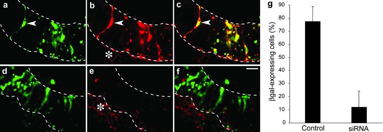
Specificity of esiRNA-mediated RNAi in the neuroepithelium of postimplantation mouse embryos developing in whole-embryo culture. E10 mouse embryos were injected into the lumen of the telencephalic neural tube with the GFP-expressing plasmid pEGFP-N2 plus the β-gal-expressing plasmid pSVpaXΔ either without (a–c and g, Control) or with (d–f and g, siRNA) β-gal-directed esiRNAs, followed by directed electroporation (lateral, cathode-right/anode-left orientation) and whole-embryo culture for 24 h. (a–f) Horizontal cryosections through the left telencenphalon were analyzed by double fluorescence for expression of GFP (green; a and d) and β-gal immunoreactivity (red; b and e). Neuroepithelial cells expressing both GFP and β-gal (arrowheads) appear yellow in the merge (c and f). Note the lack of β-gal expression in neuroepithelial cells in the presence of β-gal-directed esiRNAs. Upper and lower dashed lines indicate the luminal (apical) surface and basal border of the neuroepithelium, respectively. Asterisks in b and e indicate the basal lamina and underlying mesenchymal cells, which cross-react with the secondary antibody used to detect β-gal immunoreactivity. (Bar in f = 20 μm.) (g) Quantitation of the percentage of GFP-expressing neuroepithelial cells that also express β-gal without (Control) or with (siRNA) application of β-gal-directed esiRNAs. Data are the mean of three embryos analyzed as in a–f. (Bars indicate SD.)

