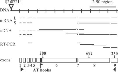
Fig 2.
The structure of the gene encoding HP2. A portion of genomic P1 clone DS00857 is shown (DNA), with the location of the 2–90 clone distal to the insertion site for P element l(2)07214. Various cDNAs and RT-PCR products sequenced to confirm the intron–exon boundaries are indicated. mRNA shows the predicted structures of the two transcripts produced by this locus (L, long; S, short). The nine exons are diagrammed below; solid triangles mark the start and stop of translation, and two solid rectangles mark the AT hooks. The locations of the genetic lesions in Su(var)2-HP2230, Su(var)2-HP2288, and Su(var)2-HP2692 also are indicated.

