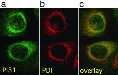
Fig 4.
(a) PI31 was visualized in B8 mouse fibroblasts by immunofluorescence staining with affinity-purified rabbit anti-PI31 antiserum and FITC-labeled secondary antibody (green). (b) The nuclear envelope/ER membrane marker PDI was visualized with monoclonal mouse anti-PDI antibody and Cy3-labeled secondary antibody (red). (c) a and b superimposed; colocalization appears in yellow.

