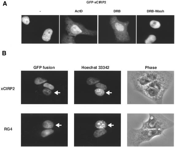
Figure 5.
Nucleocytoplasmic shuttling of xCIRP2. (A) xCIRP2 accumulates in the cytoplasm of cultured cells by treatment with transcriptional inhibitors. The GFP-xCIRP2 expression vector was transfected into HeLa S3 cells. The cells were treated with 10 µg/ml actinomycin D or 0.1 mM DRB at 37°C for 4 h, and then DRB was removed by changing the medium with fresh medium. The culture was further incubated at 37°C for 2 h. The subcellular localization of the GFP fusion proteins was analyzed by fluorescence microscopy. (B) xCIRP2 and the RG4 domain shuttle between nucleus and cytoplasm. Expression vectors of GFP-xCIRP2 (upper panels) and GFP-xRG4 (lower panels) were transfected into HeLa cells. Two days later, the cells were fused with NIH 3T3 cells to form heterokaryons and incubated in the medium containing 100 µg/ml of cycloheximide for 1 h. Heterokaryons formed between HeLa cells (human) and NIH 3T3 cells (mouse) were examined by florescence microscopy of GFP fusion proteins and nuclei stained with Hoechst 33342. The arrows indicate murine NIH 3T3 cells. ‘Phase’ panels show the phase contrast images of the heterokaryons.

