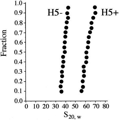
Figure 1.
Sedimentation analysis of chromatin assembled in vitro on a 6.3 kb linear DNA fragment in the absence or presence of linker histone H5. The NaCl concentration was 90 mM. Sedimentation boundaries were analyzed according to van Holde and Weischet (22). The point with boundary fraction 0.05 was omitted from the analysis because of the larger error imparted to this point by subtracting out the small constant plateau value resulting from the slow sedimenting polyglutamic acid present in the samples (see Materials and Methods). The y-axis gives the fraction of the sample that has an s20, w less than or equal to the value indicated on the x-axis.

