Figure 6.
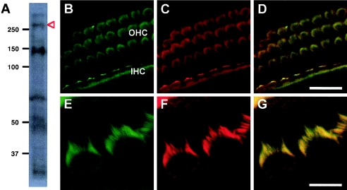
Expression and localization of TRIOBP in mouse cochlea. A, Western blot of two cochlea from newborn (P0) mice hybridized with monoclonal antibody TARA.27 (Seipel et al. 2001). Bands of ∼70 kDa and ∼50 kDa may represent two isoforms of original TRIOBP (UCSC Human Genome Browser May 2004 assembly). The band of >250 kDa (red arrow) may represent posttranslational modification of the predicted 219-kDa novel TRIOBP isoform. The band of ∼150 kDa may represent either a breakdown product of this isoform or an as-yet-uncharacterized alternatively spliced isoform. B–D, Top view of P0 whole-mount cochleae stained with monoclonal antibody TARA.27 against TRIOBP (B) and with rhodamine phalloidin, which stains F-actin (C). The protein is detected in the stereocilia of both the outer hair cells (OHC) and inner hair cells (IHC). Labeling indicates colocalization with actin along the length of the stereocilia and in the microvilli on the apical portion of the hair cells (D). E–G, Higher magnification showing stereocilia of outer hair cells. Scale bar is 16 μm in panels B–D and 4 μm in E–G.

