
Figure 6.
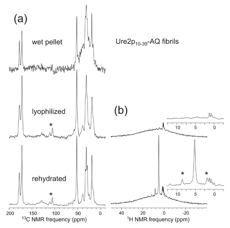
(a) 13C solid state NMR spectra of Ure2p10–39-AQ fibrils in a fully hydrated state after fibril formation (wet pellet), after lyophilization, and after rehydration. Minor changes in 13C NMR linewidths, but no changes in 13C NMR chemical shifts, are observed. Signal-to-noise for the wet pellet is lower and background signal (hump centered at 30 ppm) is higher because of the smaller sample quantity. (b) Proton NMR spectra of the lyophilized and rehydrated samples, indicating the absence of detectable mobile water (sharp peak at 5 ppm) in the lyophilized state. Asterisks indicate spinning sidebands.