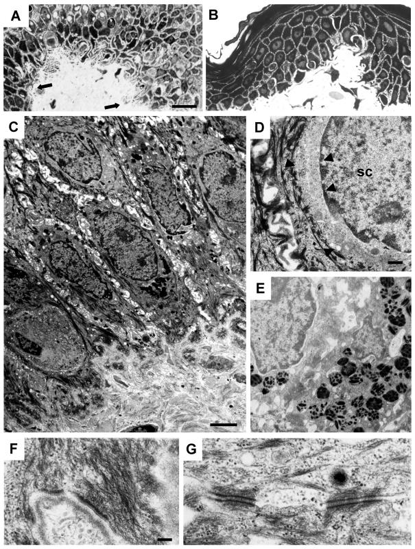
Figure 3.
Electron microscopy of skin from an individual with DDD and from a control. A, Semithin sections from a patient with DDD with filiform epithelial downgrowth (arrows), in contrast to control skin (B). Note irregular DDD cell shape and size in (A). C–G, Ultrathin sections from a patient with DDD with scattered distribution of melanosomes (E) and altered perinuclear organization of keratins in suprabasal but not in basal cells. C and D, Perinuclear rim of filament-free, smooth cytoplasm is demarcated by arrowheads; SC = spinous cell. K5 haploinsufficiency allows formation of normal keratin filaments and their interaction with hemidesmosomes (F) and desmosomes (G). Scale bars represent 20 μm (A and B), 25 μm (C), 500 nm (D and E), and 200 nm (F and G).

