
Fig. 3.
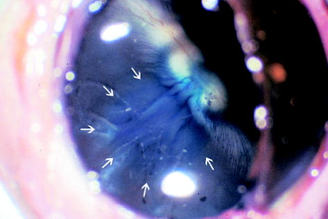
Gross picture of a rabbit eye 4 weeks after subretinal injection of the second-generation feline immunodeficiency virus vector showing the blue-stained round area (arrows) in the retina that was the retinal bleb created by the subretinal injection (original magnification, ×49.5).