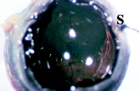
Fig. 4.

Gross picture of a rat eye 18 months after subretinal injection of the second-generation feline immunodeficiency virus vector showing the retinal bleb area stained blue (arrows). The optic nerve head and major retinal vessels are visible with normal appearance. A 12-o’clock suture (S) can be seen out of focus (original magnification, ×115).
