
Fig. 6.
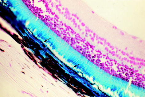
Microscopic photograph of a rat eye 24.25 months after subretinal injection of the second-generation feline immunodeficiency virus vector showing retinal pigment epithelium (RPE) cells stained blue (arrows) with normal morphology. The corresponding areas of the blue-stained retinal outer segment and choroid to the RPE staining area were obviously from diffusion of the transgene product from the RPE (original magnification, ×62.5; paraffin section, 5 μm; neutral red staining).