
Figure 1.
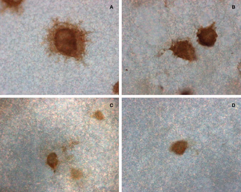
PBEF protein secretion from a representative amnion-derived WISH cell that was grown on immobilon-P membrane is shown after a 4-hour treatment with lipopolysaccharide (50 ng/mL) and immunostained to show the intracellular and secreted (extracellular) PBEF (A); there was no such halo of secreted PBEF in the control untreated cell (B). A representative primary amniotic epithelial cell was treated for 4 hours with TNF-α (30 ng/mL); the secreted PBEF was more diffuse but shown as immunostained extracellular patches (C); the extracellular staining was absent in the control (D). (Original magnification, ×400.)