FIG.4.

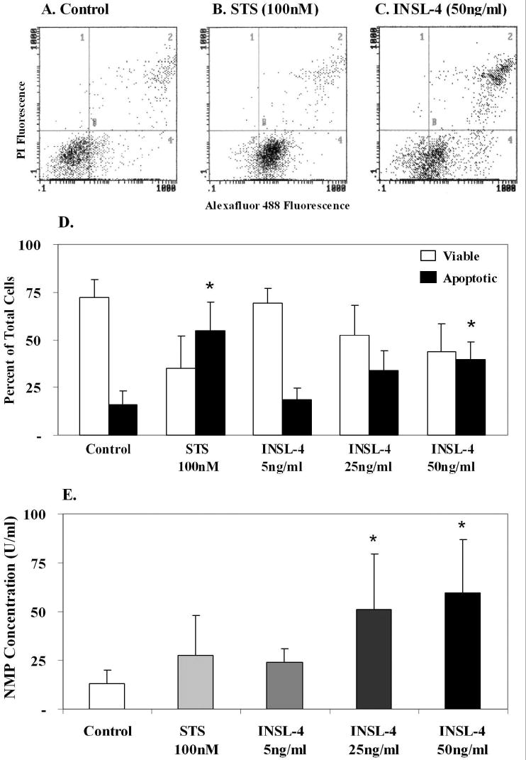
Effect of treatment of primary amniotic epithelial cells with INSL4 for 24 h (n=4, different patients). Cytograms of flow cytometric analysis: A. control, untreated cells, B. staurosporine (STS) induced apoptosis C. INSL4 treatment, showing that INSL4 also induced apoptosis and necrosis. D. quantitation of the flow cytometry data means ± SD, showing a dose-related effect of INSL4 on induction of apoptosis. Asterisks show STS significantly (p<0.05) induced apoptosis compared to the controls, INSL4 (50ng/ml) significantly induced apoptosis (p<0.05). E. Nuclear matrix protein (NMP) measurement in the medium of the experiments shown in D as means ± SD, reflect the effect of INSL4 on causing apoptosis and necrosis. Asterisks show significantly increased (p<0.05) NMP compared to the controls.
