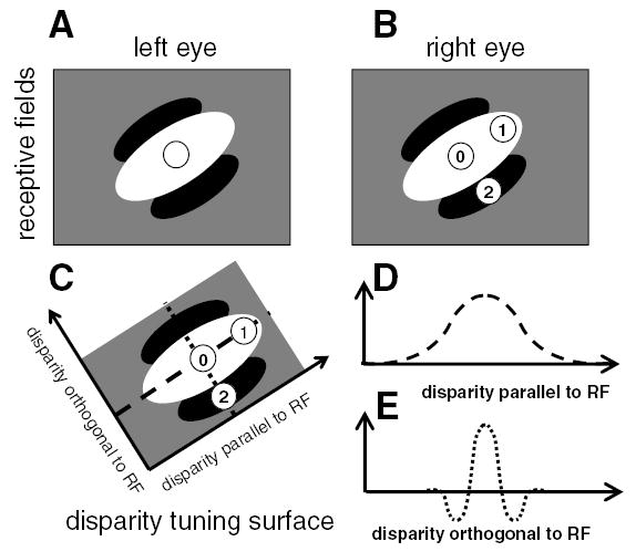
Figure 1.

Existing models predict that neurons should be more sensitive to disparities orthogonal to their preferred orientation. (A,B) The receptive fields of a binocular neuron. The left and right eye receptive fields (RFs) are identical, both consisting of a single long ON region oriented at 45 degrees to the horizontal (shown in white) and flanked by OFF regions (black). We consider stimuli with three different disparities. In each case, the left eye’s image falls in the center of the left receptive field (circle in A). The different disparities occur because the right eye’s image falls at different positions for the three stimuli. This are labeled with the numbers 0–2 in B. (C) The disparities of the three stimuli are plotted in disparity space. Stimulus 0 has zero disparity; its images fall in the middle of the central ON region of the receptive field in each eye, and so it elicits a strong response. Stimulus 1 has disparity parallel to the receptive field orientation. Although its image is displaced in the right eye, its images still fall within the ON region in both eyes, so the cell still responds strongly to both stimuli. In consequence, the disparity tuning curve showing response as a function of disparity parallel to receptive field is broad (D, representing a cross-section through the disparity tuning surface along the dashed line). Conversely, stimulus 2 has the same magnitude of disparity, but in a direction orthogonal to the receptive field orientation. Its image in the right eye falls on the OFF flank of the receptive field (B), so the binocular neuron does not respond. This leads to a much narrower disparity tuning curve; the response falls off rapidly as a function of disparity orthogonal to the receptive field (E, representing a cross-section through the disparity tuning surface along the dotted line). This means that the neuron is more sensitive to disparity orthogonal to its preferred orientation, in the sense that its response falls off more rapidly as a function of disparity in this direction.
