
Figure 6.
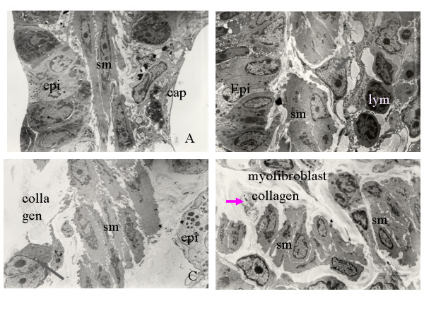
(A) Transmission electron micrograph of a bronchiole specimen from mice exposed to filtered air showing normal epithelium, smooth muscle, and capillaries. (B–D) Transmission electron micrographs of bronchiole specimens from mice exposed to 2 ppm ozone for 4, 8, and 12 weeks showing (B) hypertrophied smooth muscle cells (sm), a few infiltrating lymphocytes (lym), and myofibroblasts (arrow); (C) myofibroblasts (arrow), interstitial deposition of collagen fibers, and increased smooth muscle cell hypertrophy; and (D) disorganized smooth muscle cells, increased deposition of collagen fiber, unmyelinated nerve fiber (arrow), and myofibroblasts. Original magnification, ×5000. Scale bar = 5 μm.