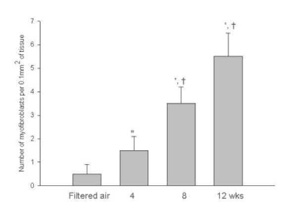
Figure 7.
Mean number of myofibroblasts counted under electron microscopy in the subepithelial zone of specimens from mice exposed to filtered air and to 2 ppm ozone for 4, 8, and 12 weeks. The myofibroblasts comprised cells with elongated projections, dilated rough endoplasmic reticulum, an infolded or crenated nuclear membrane, and bundles of parallel cytoplasmic filaments associated with dense-body condensations. * p < 0.05 compared to the group exposed to filtered air. † p < 0.05 compared to the group exposed to ozone for 4 weeks.

