
Figure 2.
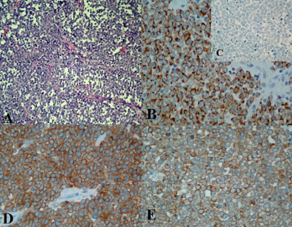
Immunohistochemical staining of primary Merkel cell carcinoma for differential diagnosis and neuroendocrine differentiation. A. Hematoxylin-eosin staining, the tumour cells have round nuclei, original magnification 200×. B. Positive cytokeratin-20 staining, showing typical punctate pattern of immunostaining, original magnification 400×. C. Negative staining for Thyroid-transcriptor factor-1, original magnification 400×. D. chromogranin -A staining, original magnification 400× and E. synaptophysin staining, original magnification 400×.