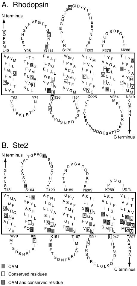
Figure 1.

Secondary structural model of rhodopsin (A) and Ste2 (B) showing the position of conserved (boxed) and constitutively active (shaded) amino acids. The transmembrane regions are enclosed in the large boxes. The topology diagrams are oriented so that the extracellular regions are shown above and the intracellular regions are shown below.
