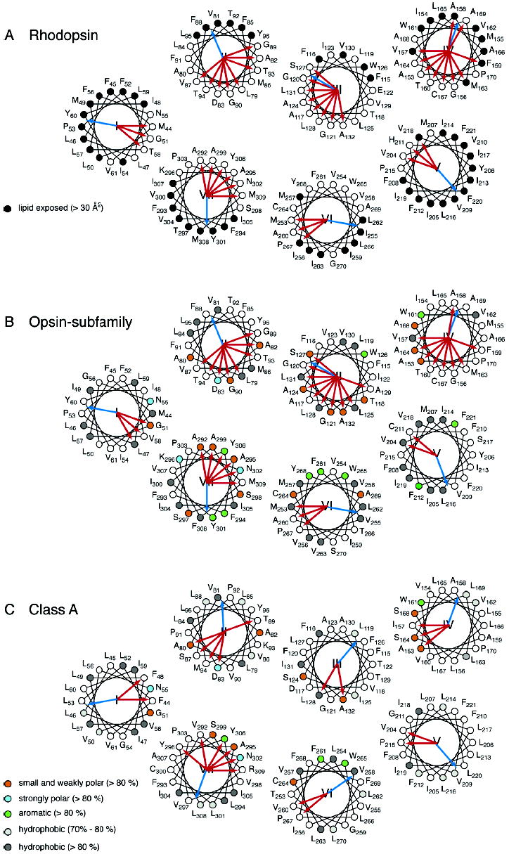
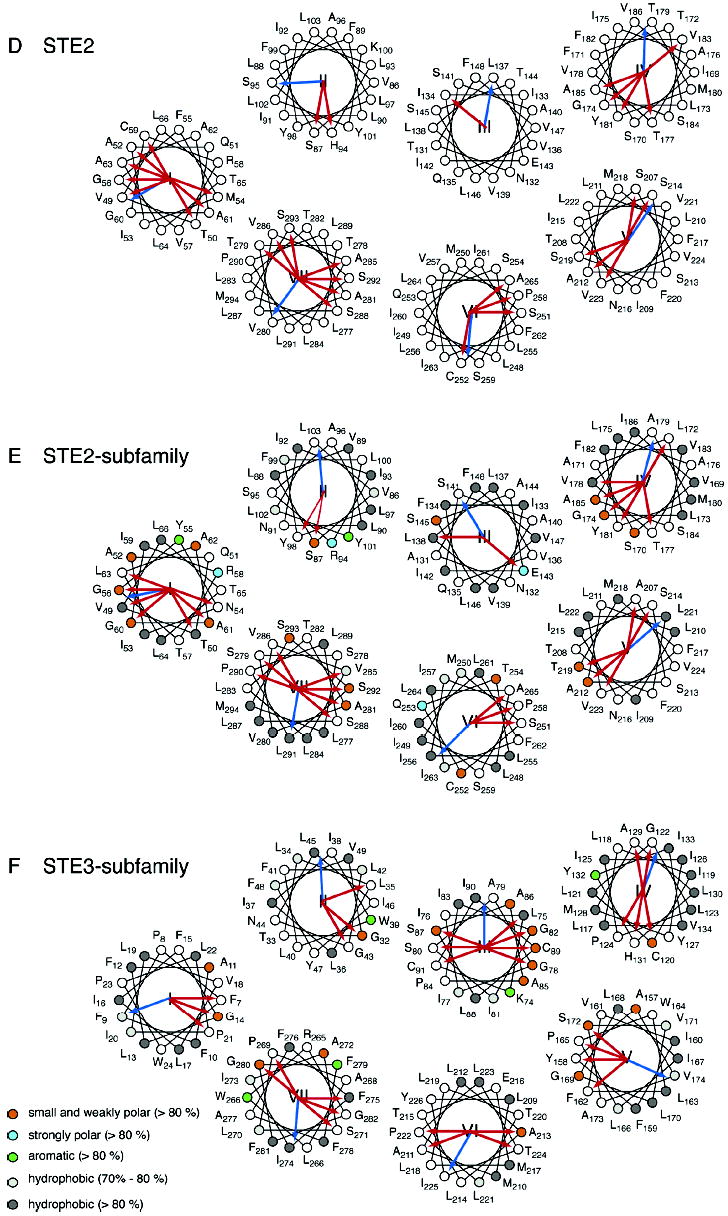
Figure 3.


Helical wheel diagrams of G protein-coupled receptors. The helical wheel diagrams show the hydrophobic moment (blue arrow) (42) and helical packing moments (red arrow) (38) for rhodopsin (A), the opsin subfamily (B), the Class A family (C), the Ste2 receptor (D), the Ste2 subfamily (E), and the Ste3 subfamily (F). For panels A–C, the positions are numbered as in bovine rhodopsin; for panels D and E, the positions are numbered as in Ste2, and in panel F, the positions are numbered as in Ste3. The sites that are group-conserved (>80%) across a family or subfamily are colored as follows: small and weakly polar (orange), strongly polar (azure), aromatic (green), and hydrophobic (dark gray). The hydrophobic sites with group conservation between 70% and 80% are light gray. For rhodopsin (A), occluded (solvent inaccessible) and exposed (solvent accessible) residues are indicated by open circles and closed circles, respectively, based on the high-resolution structure. Note that H2 in the Ste2 subfamily (D) has no helical packing moment according to our definition (38). The two strongest moments are shown as thin red arrows.
