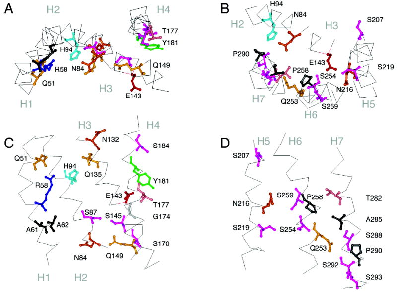
Figure 5.

Molecular model of the Ste2 receptor. Panels A and C highlight the position of several of the signature and group-conserved amino acids in helices H1 to H4. Panels B and D highlight the position of several of the signature and group-conserved amino acids in helices H5, H6, and H7. The orientations shown are the same as those in Figure 4 of rhodopsin.
