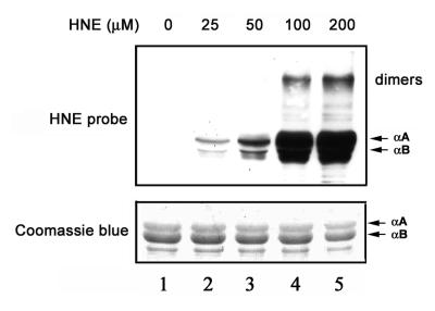
Figure 1.
Formation of HNE-protein adducts. Lane 1 is α-crystallin not treated with HNE. Lanes 2-5 are α-crystallins exposed to 25, 50, 100, and 200 μM HNE, respectively, for 2 h at 37°C. Lower panel) Protein profiles of α-crystallins isolated from bovine lenses after incubation with HNE (Coomassie Blue staining). Upper panel) modification of α-crystallins by HNE as determined by Western blotting with antibodies to HNE-modified proteins.

