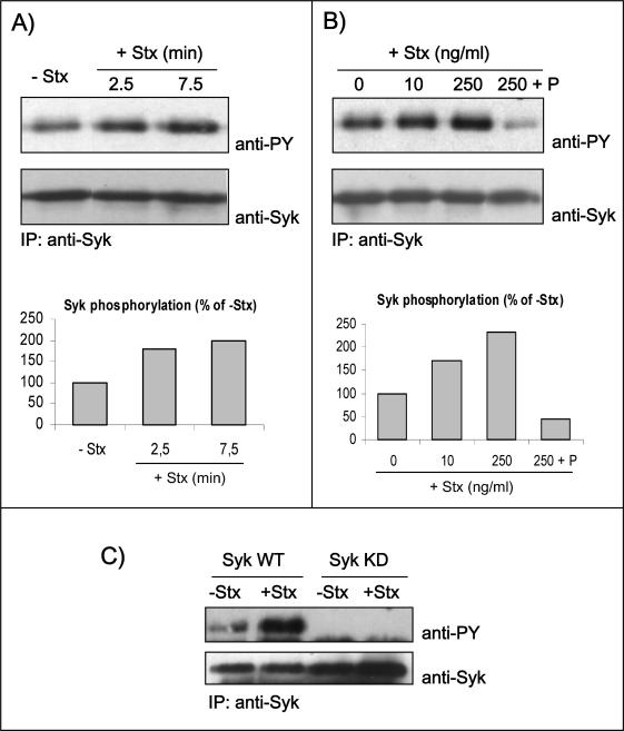
Figure 2.
Autophosphorylation of Syk in response to Stx treatment. HeLa cells transfected with human Syk cDNA and treated with 250 ng/ml Stx for 2.5 or 7.5 min at 37°C (A) or with the indicated concentrations of Stx for 5 min after preincubation with and without piceatannol (P) (50 μM) for 30 min (B). The cells were then lysed, and the lysate was immunoprecipitated overnight with anti-Syk antibody. The immunoprecipitates were analyzed by immunoblotting with mouse anti-PY. The membranes were then stripped and reprobed with mouse anti-Syk antibody. Graph, quantification of the amount of phosphorylated Syk (after normalization for the amount of immunoprecipitated Syk) in these experiments, which are representative experiments of at least three independent experiments. (C) HeLa cells transfected with cDNA encoding for Syk WT or Syk KD were incubated with and without 250 ng/ml Stx for 5 min at 37°C. Cell lysates were analyzed as described in A and B.

