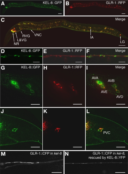
Figure 4.
KEL-8 is expressed in neurons and localized to clusters in the ventral cord dendrites. KEL-8::GFP (A, D, G, and J) and GLR-1::RFP (B, E, H, and K) fluorescence were observed throughout the entire animal (A–C), along ventral cord neurites (D–F), along lateral head ganglia (G–I), and in the lumbar ganglia of the tail (J–L). KEL-8::GFP is observed in the nerve ring (NR), lateral and ventral ganglia (L&VG), the ventral nerve cord (VNC), and the lumbar ganglia (LG) of the tail (merged image in C). Intestinal autofluorescence (IA), which is nonspecific and does not indicate expression from either transgene, is observed throughout the midbody. Along the ventral cord, KEL-8::GFP is localized to clusters adjacent to clusters of GLR-1::RFP along the ventral cord (merged image in F). KEL-8::GFP and GLR-1::RFP are expressed in the same lateral and lumbar cells bodies (merged images in I and L, respectively; cell identities are indicated). (M and N) GLR-1::CFP fluorescence was observed in kel-8 mutants (M) or kel-8 mutants (N) rescued cell autonomously with a Pglr-1::kel-8::yfp transgene containing wild-type kel-8 cDNA fused in frame to YFP. The KEL-8::YFP chimeric protein functions to rescue the kel-8 mutant phenotype in these neurons. Bars, 20 μm (A–C), 5 μm (D–L), and 10 μm (M and N).

