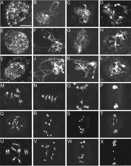
Figure 4.
Male meiosis I in the wild type and the ptd mutants. Shown are images of DAPI-stained chromosomes. (A–D) Wild-type prophase I at leptotene, zygotene, pachytene, and diplotene, respectively. (E–H) ptd-1 and (I-L) ptd-2 prophase I at similar stages showing no obvious difference to the wild type. (M–P) Wild-type diakinesis of prophase I, early metaphase I, anaphase I, and telophase I, respectively; note that M shows five brightly stained entities, representing five attached pairs of condensed homologues. (N) Chromosomes align at the equator. (O) Homologues are separating. (P) Homologues separate and form two clusters. (Q–T) Similar stages of meiosis from ptd-1. (U-X) ptd-2. Note that in Q, 10 stained bodies can be seen, indicating that the condensed homologues formed univalents. (R and V) Chromosomes condense further, similarly to normal metaphase I chromosomes, but there were no five perfectly aligned bivalents at the equator and contained univalents as in N. Chromosomes are starting to separate similar to (S and W), suggesting that they might be pulled by the spindle as normal homologues are at anaphase I. In T and X, however, the chromosome distribution seems abnormal. Bar, 10 μm.

