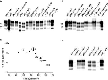
Figure 1. Determination of the Molecular Type of PrPSc Produced in Voles following Transmission of sCJD and gCJD.
(A) Immunoblot of proteinase K–resistant PrPSc from sCJD and gCJD subtypes in human patients and after first passage in voles. PrPSc produced in voles after primary transmission of mouse-passaged MM1 sCJD and MV1 sCJD, and of the mouse-adapted scrapie strain ME7 is also shown. The identity of the brain sample is designated above each lane.
(B) Comparison of proteinase K–resistant PrPSc produced in voles following first and second passages of MV1 sCJD, MM2 sCJD, E200K gCJD, and V210I gCJD. PrPSc produced in voles after first passage of the mouse-adapted scrapie strain ME7 is also shown. The identity of the brain sample is designated above each lane.
(C) Scatter-graph of proportions of di-glycosylated and mono-glycosylated PrPSc in human patients (denoted by filled circles, filled inverted triangles, filled diamonds, filled squares, and filled upright triangles) and voles (denoted by open circles, open inverted triangles, open diamonds, open squares, open upright triangles, and asterisks), with MM1 sCJD (denoted by filled squares and open squares), MV1 sCJD (denoted by filled diamonds and open diamonds), MM2 sCJD (denoted by filled circles and open circles), V210I gCJD (denoted by filled inverted triangles and open inverted triangles), E200K gCJD (denoted by filled upright triangles and open upright triangles), and ME7 (denoted by asterisks).
(D) Comparison of proteinase K–resistant PrPSc produced in voles following inoculation with MM1 sCJD, MM2 sCJD, and the mouse-adapted scrapie strain ME7. Strep-tagged molecular markers (25 kDa and 20 kDa) are shown. The identity of the brain samples is designated above each lane.

