
Figure 5.
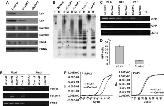
Silencing of Lsh in embryonic stem cells results in loss of de novo methylation. (A) Western analysis: nuclear extracts from two ES cell lines (#1 and #2 both received the same construct) stably expressing a silencing vector for Lsh (siLsh) were examined by Western analysis using specific antibodies against Lsh, Dnmt3a, Dnmt3b, Chd4 or PCNA as control. A scrambled sequence serves as the siRNA vector control. (B) Southern blot analysis: genomic DNA derived from two siLsh ES cell lines, an ES cell control, Lsh−/− MEFs, and Lsh+/+ MEF cells was digested with HpaII (H) or MspI (M), blotted, and probed for minor satellite sequence using the probe MR150. (C) RT–PCR analysis: siLsh ES cells were infected with the retroviral vector pMSCV-hGFP and after the indicated time points (24, 48, 72 h) RNA was extracted, reverse transcribed and analyzed by PCR for expression of GFP. β-Actin serves as a control. (D) FACS analysis: 72 h after retroviral infection of siLsh and control ES cells GFP expression was measured by FACS analysis. (E) Methylation-sensitive PCR: genomic DNA derived from the siLsh and control ES cells 3 days after retroviral infection was digested with HpaII or MspI and subjected to PCR analysis with the indicated primer pairs. (F) Real time-PCR analysis: as in (E), HpaII digested genomic DNA was subjected to real time-PCR using P11/P12. The right panel shows the internal control reaction with primers P7/P8.