
Figure 3.
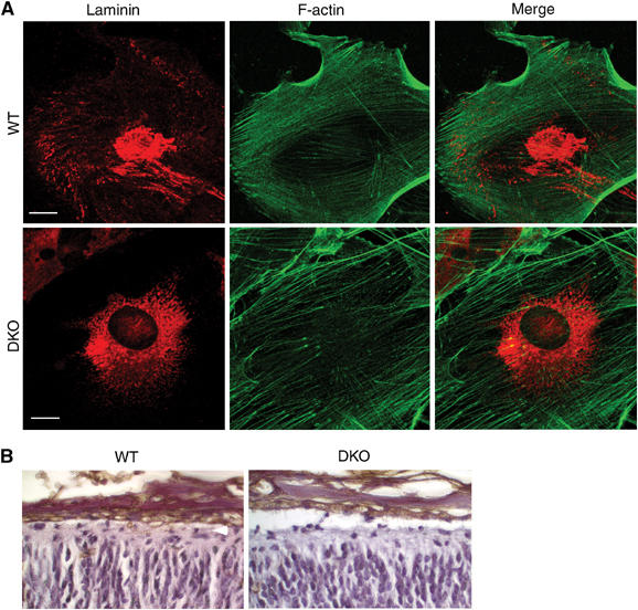
Defective laminin organization in FE65−/−; FE65L1−/− meningeal fibroblasts. (A) Primary meningeal fibroblasts from WT and FE65−/−; FE65L1−/− mice were double stained with a laminin antibody and phalloidin-AlexaFluor 488 to detect F-actin. Fibrillar and punctate laminin is observed at the periphery in WT meningeal fibroblasts, whereas it is absent from FE65−/−; FE65L1−/− meningeal fibroblasts. Scale bar, 20 μm. (B) Staining with the lectin RCA-1 shows no difference in the thickness of meninges for WT and FE65−/−; FE65L1−/− E18.5 mouse brains.