
Figure 4.
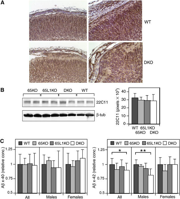
APP and Aβ levels in FE65−/−; FE65L1−/− brains. (A) IHC staining for APP (A8717) shows expression throughout the cortical plate in WT and FE65−/−; FE65L1−/− E18.5 brains and in heterotopic cells. (B) APP and APLP2 steady-state levels are unchanged in FE65−/−; FE65L1−/− adult mouse brains compared to WT. Western blot analyses using 22C11 and anti-β-tubulin (reprobe) on two mouse brain diethylamine extracts per genotype. Bar chart shows average APP steady-state levels that were normalized to β-tubulin (as load control), with standard error bars for WT (n=8), FE65−/− (n=7), FE65L1−/− (n=9), FE65−/−; FE65L1−/− (n=9) mouse brains. No significant difference was observed between genotypes. (C) Aβx-40 and Aβx-42 levels in diethylamine brain extracts of WT (n=5F, 7M), FE65−/−(n=7F, 4M), FE65L1−/− (n=10F, 4M) and FE65−/−; FE65L1−/− (n=6F, 6M) 14- to 21-week-old mice. Data have been normalized to the mean of the WT mice. Aβx-42 levels are significantly reduced in FE65−/−; FE65L1−/− males compared to WT. t-Test *P<0.05; **P<0.005.