Figure 3.

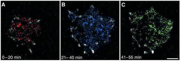
Reorganization of QDs under spreading cells. The area under the cells that is covered with hyaluronan is indicated by the history of QDs translational movements in the XY plane. Each appearance of a QD is represented by a colored pixel. All pixels are plotted in a cumulative manner such that all the coordinates in which QDs appeared are marked with a colored pixel. Paxillin-containing structures are white. (A) During the first 20 min, QDs appear all over the cell-surface interface; (B) 21–40 min after seeding, additional QDs accumulate under the cell, displaying an increasingly nonuniform distribution. (C) As the cell continues to spread (41–55 min), QDs becomes confined to discrete areas under the cell, in the area located between focal adhesions, and their movement in the XY plane become restrained. Scale bar=5 μm.
