
Figure 4.
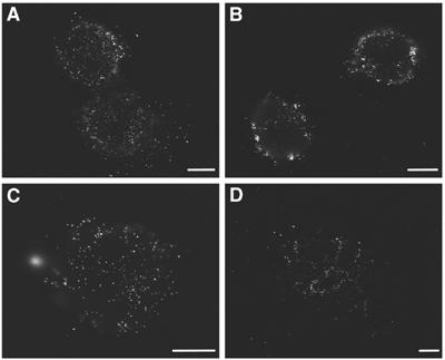
Reorganization of hyaluronan under the cell during spreading. Hyaluronan reorganization is observed by scanning electron microscopy using QX™ capsules. RCJ-P cells were labeled with biotinylated hyaluronan-binding protein and streptavidin-conjugated colloidal gold, and seeded in a QX™ capsules for 5, 10, 20, and 30 min before fixation. Gold particles are visible on the ventral side of the cell membrane (white particles). (A) Gold particles are scattered evenly under the cells 5 min after seeding. (B) After 10 min the gold particles appear more organized within discrete areas under the cells. (C, D) At 20 and 30 min after seeding, the gold particles become increasingly clustered in compartments under the cells. Scale bars=5 μm.