
Figure 5.
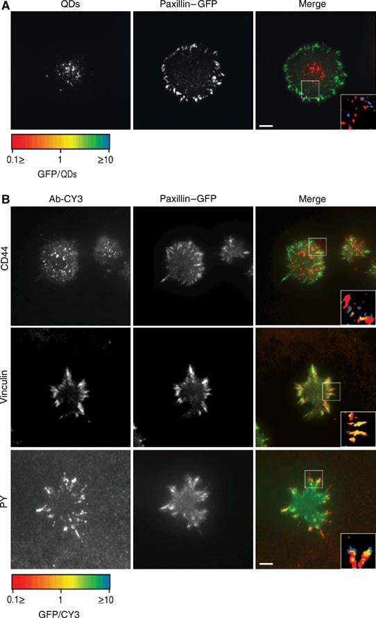
Hyaluronan-bound QDs and CD44 do not colocalize with paxillin-containing structures. (A) RCJ-P chondrocytes stably expressing paxillin–GFP were labeled with biotinylated hyaluronan-binding protein and streptavidin-conjugated QDs, seeded on fibronectin-coated glass (10 μg/ml) and incubated for 20 min before fixation. Left: QDs; middle: paxillin–GFP; right: merged image. (B) RCJ-P chondrocytes stably expressing paxillin–GFP were seeded on uncoated glass, incubated for 20 min, fixed and immunolabeled for CD44, vinculin and phosphotyrosine. Left: antibody-CY3 expression; middle: paxillin–GFP; right: merged image. Insets: fluorescence ratio analysis of the areas marked with a box. Red: structures with high paxillin–GFP/Ab-CY3 or paxillin–GFP/QDs ratio, Blue: structures with low paxillin–GFP/Ab-CY3 or paxillin–GFP/QDs ratios. Yellow: paxillin–GFP/Ab-CY3 or paxillin–GFP/QDs, normalized ratio=1. Scale bar=5 μm. (A, B) The localization of QDs and of the hyaluronan receptor CD44 is negatively correlated with paxillin–GFP. (B) Vinculin colocalizes with paxillin,phosphotyrosine colocalizes with the tips of paxillin-containing structures.