
Figure 2.
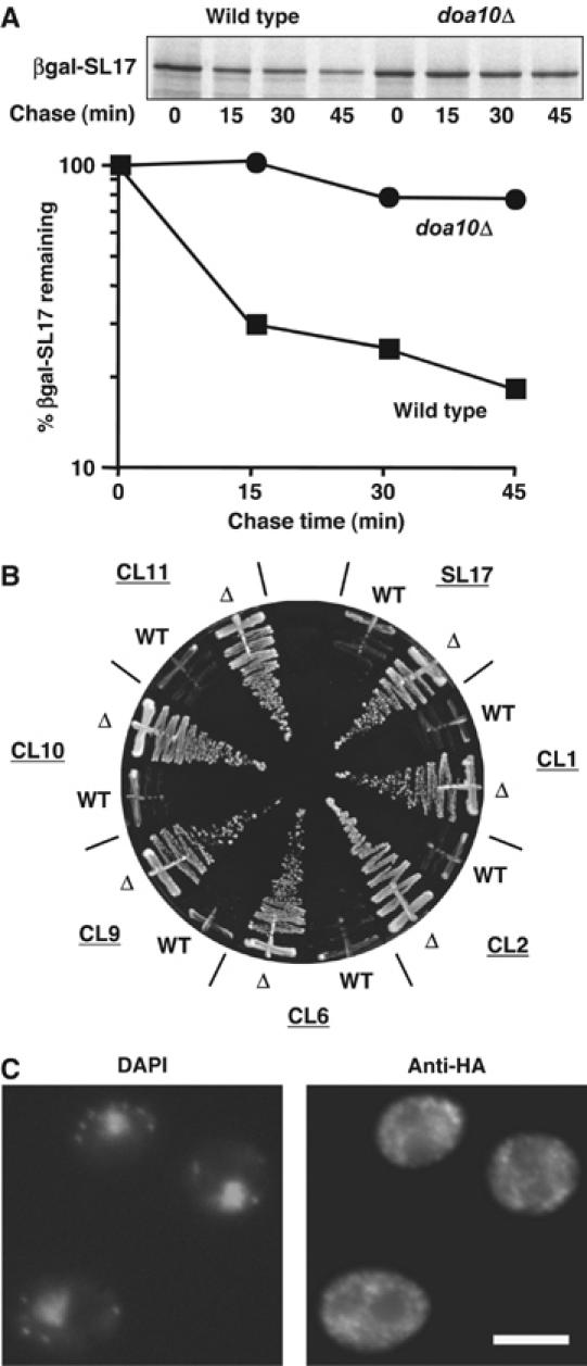
Doa10 substrates include a series of cytoplasmic synthetic degron fusions. (A) Degradation of a fusion of the SL17 degron with βgal measured by pulse-chase analysis at 30°C. (B) Growth on SD-ura of wild-type (WT) and doa10Δ (Δ) cells expressing fusions of the indicated degrons to Ura3. (C) Cytoplasmic localization of Ura3-HA-SL17 detected by anti-HA immunofluorescent staining in doa10Δ. Staining was similar in WT and doa10Δ cells, but much brighter in the latter; three fusions (SL17, CL1 and CL2) were tested and gave similar results. Scale bar, 5 μm.