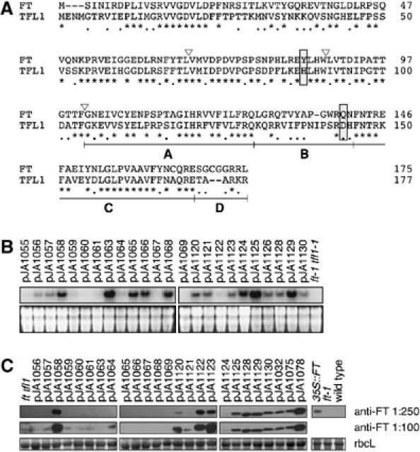
Figure 1.

Sequence and expression of chimeras. (A) Amino-acid sequences of the parental FT and TFL1 proteins. Asterisks indicate identical residues, dots residues with similar biochemical properties, and dashes gaps introduced to optimize the sequence alignment. Triangles show the exon boundaries of FT and TFL1. The four segments used to generate the segmental chimeras within the fourth exon are shown as A, B, C and D. The Tyr85/His88 and Gln140/Asp144 residues, which form a hydrogen bond in TFL1, but not FT, and which are likely the most critical residues for distinguishing FT and TFL1 activity, are boxed. (B) RNA accumulation in transgenic plants. Blots were probed with a mixture of FT and TFL1 probes. The bottom panels show ethidium bromide-stained gels as loading controls. (C) Protein blot analysis using anti-FT antibody. The bottom panels show Ponceau S-stained blots (major band is large subunit of Rubisco).
