
Figure 1.
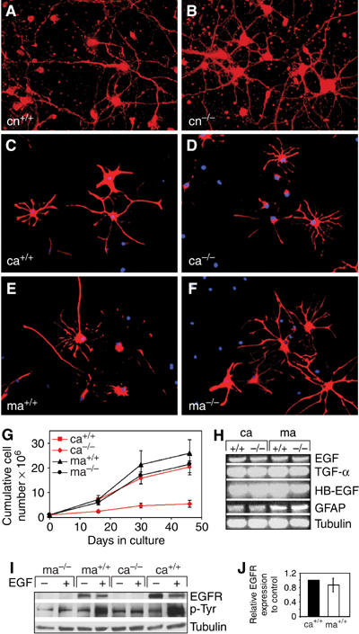
The EGFR is dispensable in neurons but differently required in astrocytes from various brain regions. (A, B) Cultured EGFR+/+ (cn+/+) and EGFR−/− (cn−/−) cortical neurons stained for the neuronal marker GAP-43 (red). (C–F) Immunofluorescence staining for GFAP (red) of cultured cortical EGFR+/+ (ca+/+) and EGFR−/− (ca−/−) and midbrain EGFR+/+ (ma+/+) and EGFR−/− (ma−/−) astrocytes. DAPI (blue) was used as a nuclear counterstain. (G) Cumulative cell numbers of EGFR+/+ and EGFR−/− cortical and midbrain astrocytes. Data represent mean±s.e.m. of four independent experiments. (H) RT–PCR analysis showing expression of GFAP and of the EGFR ligands EGF, TGFα and HB-EGF. Tubulin is used as loading control. (I) Western Blot analysis showing EGFR protein expression and tyrosine phosphorylation after stimulation with 50 ng/ml EGF. Tubulin is used as loading control. (J) Quantification of EGFR protein levels present in cortical and midbrain astrocytes relative to tubulin. Data represent mean±s.e.m. of three independent Western blot analyses normalized to control.