
Figure 6.
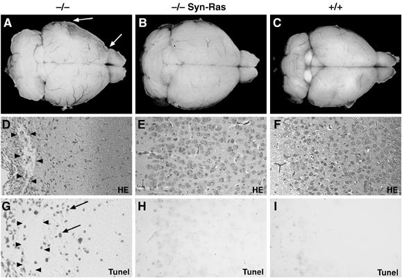
Expression of RasV12 in neurons rescues the brain degeneration in EGFR−/− mice. Dorsal view of the whole brain of (A) EGFR−/− mice with cortical degeneration (arrows), (B) EGFR−/− mice expressing the RasV12 transgene in postmitotic neurons (−/− Syn-Ras) and (C) control mice isolated at postnatal day 16. (D–F) Histological sections showing extensive degeneration evidenced by large cysts (arrowheads) in EGFR−/− cortex (D), and normal architecture in EGFR−/− Syn-Ras (E) and wild-type littermate control cortex (F). (G–I) Tunel staining showing apoptotic neurons in EGFR−/− cortex (arrows) (G) but not in EGFR−/− Syn-Ras (H) and control cortex (I). Magnification: D–I, × 20.