
Figure 7.
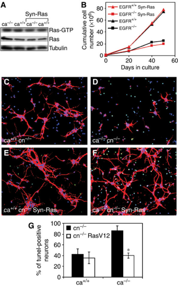
EGFR−/− Syn-Ras transgenic neurons survive in co-cultures with EGFR−/− astrocytes. (A) Western blot analysis showing levels of Ras-GTP in cultured astrocytes of the indicated genotypes. (B) Cumulative cell number of primary astrocytes demonstrating that EGFR−/− Syn-Ras transgenic astrocytes display a growth defect comparable to EGFR−/− astrocytes. (C–F) Cortical EGFR−/− (C, D) and EGFR−/− Syn-Ras (E, F) neurons (cn) co-cultured with wild-type (C, E) and EGFR−/− (D, F) cortical astrocytes (ca). Immunofluorescence stainings were performed 12 days after co-culture using GAP-43 (red) as neuronal marker, DAPI (blue) as a nuclear counterstain and Tunel (green) for apoptotic neurons. (G) The percentage of Tunel-positive neurons present in the indicated co-cultures at day 12 is shown. Data represent the mean±s.e.m. of the number of apoptotic cells counted in randomly chosen fields of six independent samples. *P<0.05.