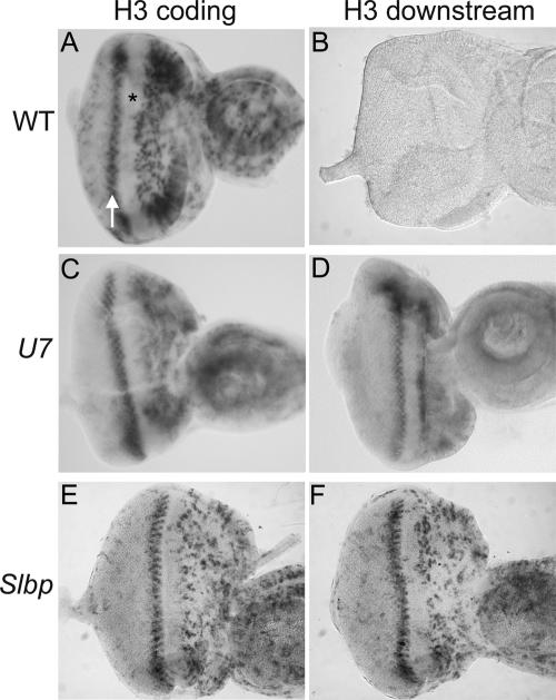
FIGURE 5.
U7 mutant eye imaginal discs display a wild-type pattern of polyadenylated H3 mRNA. Eye imaginal discs were dissected from w1118 control (A,B), U714 (C,D), or Slbp15 (E,F) mutant larvae and subjected to in situ hybridization with an H3 coding probe (A,C,E) or the H3-ds probe (B,D,F), which detects only misprocessed, poly A+ H3 mRNA. The asterisk indicates the morphogenetic furrow, which contains cells arrested in G1 phase. The arrow indicates S phase of the second mitotic wave. Asynchronously dividing, undifferentiated cells are located anterior of the MF (right of the asterisk), and differentiating cells that have exited the cell cycle and that will make up the adult eye are located posterior to the MF (left of the arrow).

