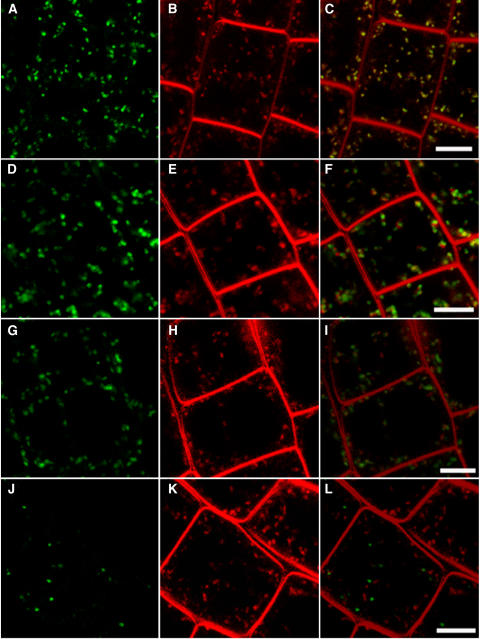
Figure 3.
Rapid Colocalization of VHA-a1–GFP with FM4-64.
Cells in the root elongation zone expressing different GFP markers were stained with the endocytic tracer FM4-64: VHA-a1–GFP (A), N-ST-YFP (D), ARA7-GFP (G), and ARA6-GFP (J). Overlays of the separately recorded GFP (green) and FM4-64 (red) channels ([B], [E], [H], and [K]) are shown in (C), (F), (I), and (L). All images were taken after 6 min of FM4-64 uptake. Bars = 10 μm.

