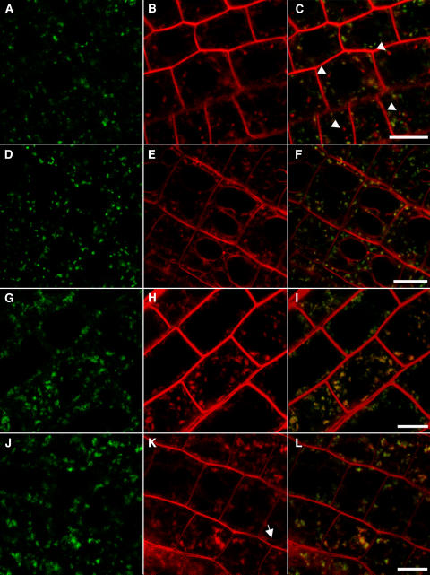
Figure 4.
ConcA Blocks FM4-64 Transport to the Tonoplast.
Cells in the root elongation zone expressing VHA-a1–GFP were stained with the endocytic tracer FM4-64. Shown are the GFP channel ([A], [D], [G], and [J]), the FM4-64 channel ([B], [E], [H], and [K]), and overlays of the separately recorded GFP (green) and FM4-64 (red) channels ([C], [F], [I], and [L]). Bars = 10 μm.
(A) to (C) Untreated cells expressing VHA-a1–GFP 1 h after FM4-64 staining. Arrowheads mark FM4-64 staining separate from VHA-a1–GFP signal that might represent later endosomal compartments.
(D) to (F) Untreated cells expressing VHA-a1–GFP 2 h after FM4-64 staining.
(G) to (I) ConcA-treated seedling root cells expressing VHA-a1–GFP 2 h after FM4-64 staining. Note the absence of FM4-64 staining not associated with VHA-a1–GFP and of tonoplast staining.
(J) to (L) ConcA-treated seedling root cells expressing VHA-a1–GFP 5 h after FM4-64 staining. The arrow indicates faint tonoplast staining.

