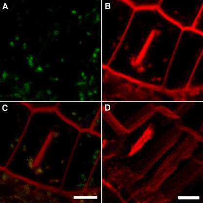
Figure 7.
Nascent Cell Plates Are Rapidly Stained by FM4-64.
(A) to (C) Dividing cells expressing VHA-a1–GFP were stained with the endocytic tracer FM4-64. Images were taken within 15 min of FM4-64 staining. Shown are the separately recorded GFP (A) and FM4-64 (B) channels as well as the overlay (C).
(D) Three-dimensional reconstruction of an image stack derived from a Z scan shows that the cell plate is not connected to the surrounding plasma membrane.
Bars = 5 μm.

