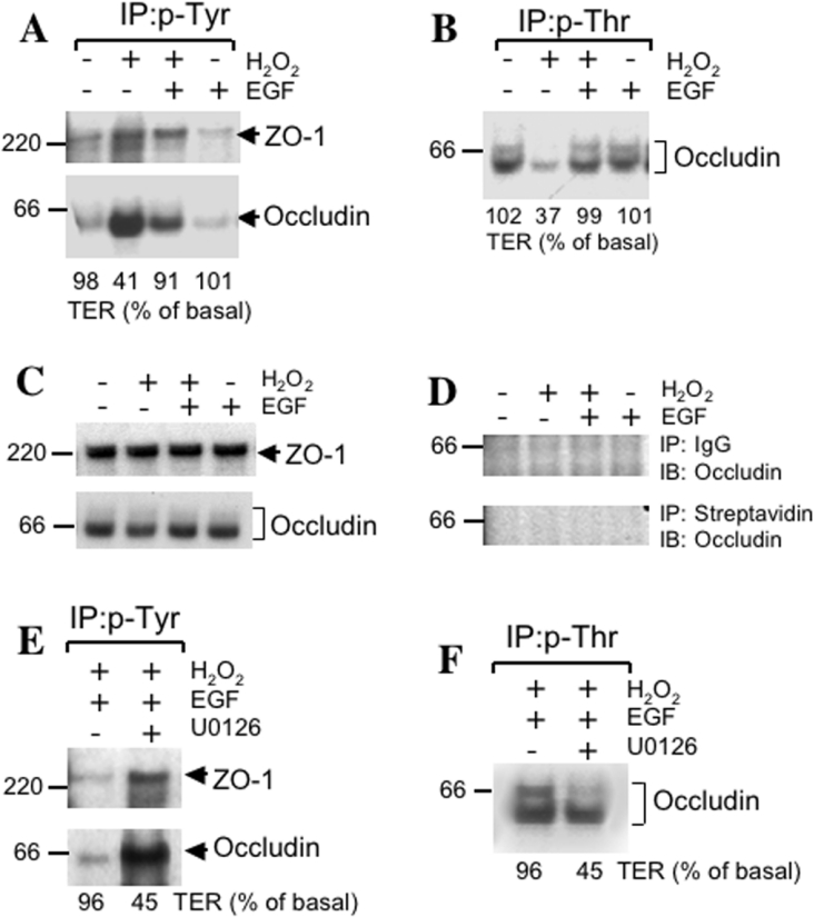
Figure 2. EGF prevents H2O2-induced alteration in phosphorylation of occludin and ZO-1 by a MAPK-dependent mechanism.
(A) and (B) Caco-2 cell monolayers were incubated with or without H2O2 in the presence or absence of 30 nM EGF for 3 h. Proteins extracted under denatured conditions were subjected to immunoprecipitation of p-Tyr (A) or p-Thr (B) followed by immunoblot analysis for occludin and ZO-1. Values at the bottom of immunoblots represent the TER values for the corresponding experiment. This experiment was repeated twice with similar results. (C) and (D) Caco-2 cell monolayers were incubated as described above for (A) and (B). Tissue extracts were either directly immunoblotted for total occludin and ZO-1 (C) or control-immunoprecipitation was performed using preimmune rabbit-IgG with Protein A–Sepharose or streptavidin–agarose followed by immunoblot analysis for occludin and ZO-1. (E) and (F) Caco-2 cell monolayers were incubated with EGF and H2O2 in the presence or absence of U0126 for 3 h. Extracted proteins were subjected to immunoprecipitation of p-Tyr (E) or p-Thr (F) followed by immunoblot analysis for occludin and ZO-1. This experiment was repeated twice with similar results.

