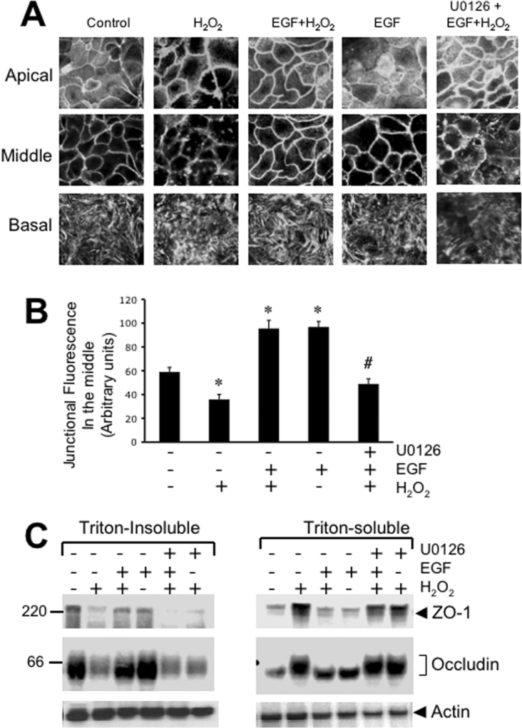
Figure 3. EGF prevents H2O2-induced reorganization of actin cytoskeleton and its interaction with TJ proteins by a MAPK-dependent mechanism.
Caco-2 cell monolayers were incubated with or without H2O2 in the presence or absence of EGF and U0126 for 3 h. (A) Cell monolayers were fixed in paraformaldehyde and stained with Alexa Fluor™ 488-conjugated phalloidin. Fluorescence images from different depths in epithelium (apical, middle and basal) were obtained by confocal microscopy. (B) Semi-quantitative analysis of junctional fluorescence in the middle part of the cells. Intensity of fluorescence at the junctions was measured over a constant area of 1 mm2 and the values are presented as arbitrary units. Values are mean±S.E.M. (n=4; each value is the average of 4 different regions from the same image). *, indicate the values that are significantly different (P<0.05) from values for control cells; # indicates the values that are significantly different from values for the EGF+H2O2 group. (C) Triton-insoluble and Triton-soluble fractions were prepared from different cell monolayers and immunoblotted for occludin, ZO-1 and actin.

