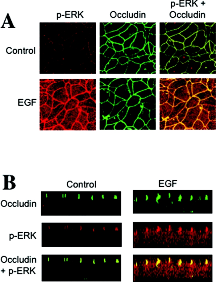
Figure 5. p-ERK is co-localized with occludin at the TJ in Caco-2 cell monolayers.
(A) Caco-2 cell monolayers were incubated in the presence or absence of EGF for 5 min. Cell monolayers were fixed in acetone/methanol and stained for occludin and p-ERK by immunofluorescence methods. (B) Optical z-sections of fluorescence images were collected by confocal microscopy.

