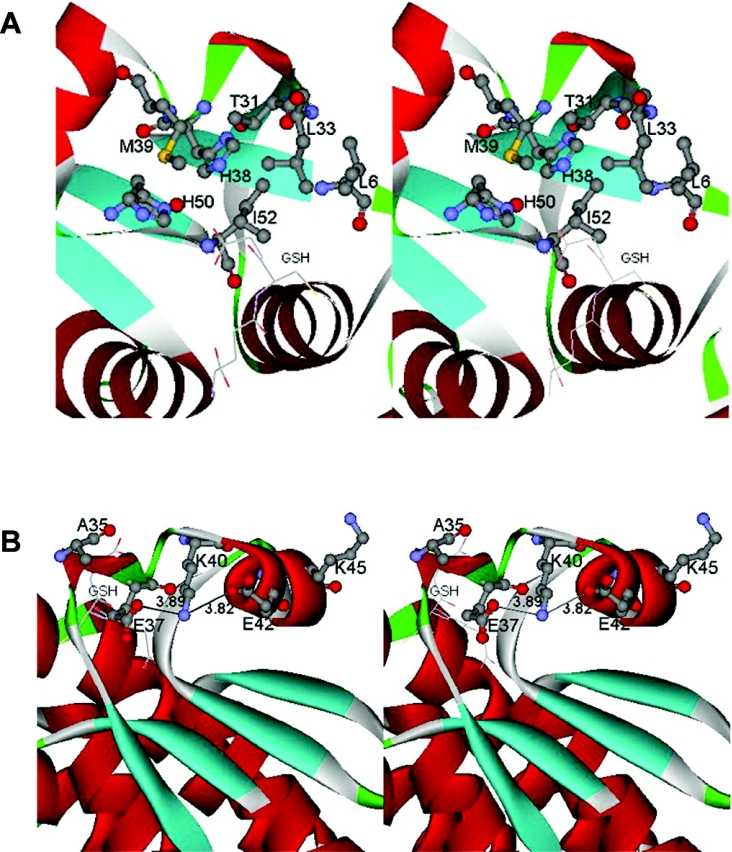
Figure 1. Stereo views of domain I region characterized in this study.
(A) Stereo view of the active-site pocket of adGSTD4-4. Leu-6, Leu-33, Thr-31 and Ile-52 form part of the G-site wall. The hydrophobic residues Leu-6, Leu-33 and Ile-52 form a small hydrophobic cluster in the G-site. GSH in the active site is shown in stick form. (B) Stereo view of the charged residues located near the α2 helix of adGSTD4-4. The ionic bridge motif formed by the charged residues (Glu-37, Lys-40 and Glu-42) is exposed to the solvent, located at the N-terminus of the α2 helix. GSH in the active site is shown in stick form. Both parts of the Figure were created with Accelrys DS ViewerPro 5.0.

